
sphenopalatine sinus horse
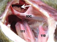
Caudal Maxillary Sinus 3. To describe a novel standing trans-nasal endoscopic guided CO 2 laser fenestration approach to access the sphenopalatine sinus SPS in the horse.

Die Anatomie Der Haustiere Veterinar Anatomie Die Nasennebenhohlen 85 Superior Kieferhohle Ist Auch Bj Den Infra Orbital Kanal Uber Den Es Freeh Offnet In Die Sphenopalatine Sinus Gekreuzt Es Connnunicates Dorsal Mit Dem
The sphenoidal and palatine sinuses communicated in most horses.

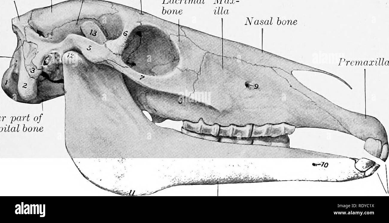
. Knowledge of the paranasal sinus anatomy is the most important aspect of the procedure. This sinus lies under the ethmoidal labrynth. However the clinical anatomy of these sinuses is not well described in.
The cribriform plate and osseous structures of the sphenopalatine sinuses appeared intact Fig. They extend down the face to the lower end of the cheekbones. ArticleBertuglia2006SphenopalatineSS titleSphenopalatine sinus syndrome in a horse authorAndrea Bertuglia and Antonella Rampazzo and A Brignolo and Antonio Dangelo journalIppologia year2006 volume17 pages13-16.
Cadaver study and client-owned 20-year-old Warmblood gelding. Rostral Maxillary Sinus 2. Gas attenuation surrounded the roots of tooth 210 and sclerosis and blunting of these roots were additionally observed.
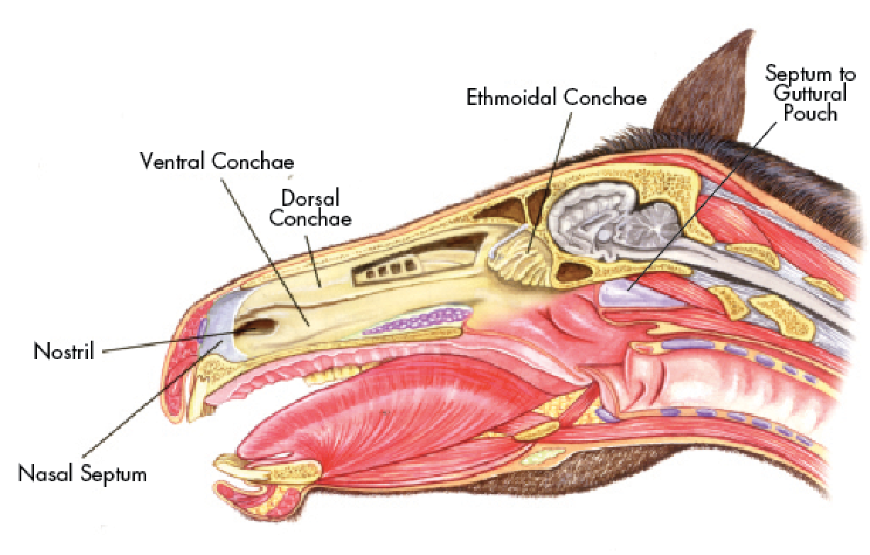
Clinical anatomy of the equine sphenopalatine sinus McCANN J. It is divided into rostral and caudal compartments by an oblique bony septum that continues dorsally into a thin domed structure the maxillary septal bulla formerly incorrectly termed the ventral conchal bulla. There are 6 paired sinuses frontal maxillary dorsal conchal ventral conchal middle conchal and sphenopalatine and all of these spaces communicate with each other and the nasal passage either directly or indirectly.
The maxillary sinus is the largest of the sinuses. Cattle do not have a fully developed sinus system until the age of 7 by which time it is an extremely complex system. Sinoscopy is a minimally invasive diagnostic procedure that can be performed on the standing horse.
The sphenopalatine sinus drains via the caudal maxillary sinus with which is communicates freely over the infraorbital canal. Often referred to as paranasal sinuses because they are near the nose sinuses have a smooth interior lining and are covered by a thin layer of bone. Sphenopalatine sinus syndrome in a horse.
Request PDF Sphenopalatine sinus syndrome in a horse A 14-years-old Arabian mare presented for clinical consultation with a 2-months history of chronic nasal discharge and recurrent epistaxis. The aim of this study was to produce an anatomical atlas to support computed tomography CT and sinuscopy of the paranasal. Use of smaller diameter or specialized instruments such as various endoscopic bone cutting instruments and CT image guidance may.
In terms of management lavage flushing out is key García-López said. Nickels Henry ONeill in Equine Surgery Fifth Edition 2019 Maxillary Sinus. Ventral Conchal Conchal Sinus 4.
2004-01-01 000000 Reasons for performing study. Dorsal Conchal Sinus 9. No abnormalities associated with the 109 tooth extraction site were detected.
The incision for the portal into the caudal maxillary sinus is centered 2 cm rostral and 2 cm ventral to the medial canthus of the eye. The horses head has uniquely adapted itself and developed six pairs of paranasal sinusesthe frontal sphenopalatine and maxillary sinuses and the dorsal middle and ventral conchal sinuses. The technique can allow good visualization and treatment of paranasal sinus disease and eliminate the need for more invasive procedures such as large bone flaps.
Clinical and radiographic investigations of paranasal sinuses in horses are difficult due to the complex anatomy of these regions the lack of patognomonic symptoms and the low sensitivity of conventional diagnostic techniques. The same structures observed through the portal in the frontal bone can be identified directly through this portal but in some instances the entrance to the sphenopalatine sinus is inaccessible. The equine paranasal sinuses PNS are an.
Anatomy of the Horses Sinuses 1. Sphenopalatine Sinus In the horse the sphenoid and palatine sinus compartments communicate and are hence known as the sphenopalatine sinus. Inspissated thickened pus accumulated in this horses sinuses especially in the ventral conchal sinus.
In such cases what could accurately be termed the combined sphenopalatine sinuses usually drained directly into the caudal. Transnasal endoscopically-guided ventral surgical access to the sphenopalatine sinus is possible in horses and may improve access in horses with disease extending caudally beyond the palatine portion of the sinus. Disorders of the equine sphenopalatine sinus including empyema and neoplasia have been reported to cause damage to cranial nerves II and V.
In addition the horse has sphenopalatine and ethmoidal sinuses which are of a lesser clinical importance than the frontal caudal maxillary and rostral maxillary sinuses on each side of the skull. The maxillary sinus is the largest paranasal sinus and is divided into two parts rostral and caudal by a thin septum. Sinuses are air-filled cavities located on either side of the horses head above below and between the eyes.
Septum between Maxillary Sinuses 11. The caudal maxillary sinus Sinus maxillaris caudalis SMC is the central compartment of the caudal paranasal sinus system and opens into the dorsal conchal sinus Sinus conchae dorsalis SCD the middle conchal sinus Sinus conchae mediae SCM the frontal sinus Sinus frontalis SF and the sphenopalatine sinus Sinus sphenopalatinus SSP.

Equine Cardiovascular Respiratory Flashcards Quizlet

Equine Head Special Structures Of The Paranasal Sinuses Flashcards Quizlet
2

Researchers Cast New Light On Equine Sinuses Horsetalk Co Nz

Equine Airway Issues Springhill Equine Veterinary Clinic
![]()
Transverse Ct Images Of Head Of A Horse With Right Sided Sinusitis Download Scientific Diagram

Pdf Bit Induced Asphyxia In The Horse Elevation And Dorsal Displacement Of The Soft Palate At Exercise Semantic Scholar
Paranasal Sinus Anatomy And Trephination Technique Proceedings

Direct Approach To The Nasal Cavity Through A Bone Flap For The Treatment Of A Large Nasal Cyst Bolz 2017 Equine Veterinary Education Wiley Online Library
Equine Sinus Disease A Hidden Danger

Horses With Facial Swelling Nasal Discharge Flashcards Quizlet

Equine Nasal Discharge Flashcards Quizlet
Equine Sinus Conditions Large Animal Hospital College Of Veterinary Medicine University Of Florida
Equine Paranasal Sinus Disease Questions Facial Swelling And Nasal Discharge Flashcards Quizlet

Equine Nasal Discharge Flashcards Quizlet

Die Anatomie Der Haustiere Veterinar Anatomie Die Nasennebenhohlen 85 Superior Kieferhohle Wird Auch Durch Die Infra Orbital Kanal In Dem Er Sich Frei In Die Sphenopalatine Sinus Offnet Sich Gekreuzt Es Kommuniziert Dorsal
Die Anatomie Der Haustiere Veterinar Anatomie Die Nasennebenhohlen 85 Superior Kieferhohle Wird Auch Durch Die Infra Orbital Kanal In Dem Er Sich Frei In Die Sphenopalatine Sinus Offnet Sich Gekreuzt Es Kommuniziert Dorsal

Computed Tomographic Images Of The Equine Head Region The Patient S Download Scientific Diagram

Standing Trans Nasal Endoscopic Guided Co2 Laser Fenestration Of The Palatine Bone To Access The Sphenopalatine Sinus In A Horse Perez 2021 Veterinary Surgery Wiley Online Library


0 Response to "sphenopalatine sinus horse"
Post a Comment